Main Text
1 Introduction
Esophageal cancer is a common and highly invasive gastrointestinal tumor, which is caused by pathological changes of esophageal epithelial mucosa cells, with difficulty in eating and dysphagia as the main manifestation, seriously affecting the quality of patients' life, with a high incidence and mortality [1-4]. Since the tumor of early esophageal cancer only invades the mucosa or submucosa instead of the deep layer, without the occurrence of lymph node metastasis and the early symptoms, it is often difficult for patients to detect. This disease can easily progress to the middle and late stages, which increases the difficulty of treatment, eventually leading to poor prognosis. Therefore, accurate diagnosis and intervention in the early stages of the disease are effective means of reducing the incidence and mortality of esophageal cancer, which is important for improving the clinical outcome of patients with esophageal cancer [5,6].
Gastroscopic biopsy is the current gold standard for the diagnosis of esophageal cancer, but it is more invasive to the patient, which leads to poor compliance [7,8]. Three-dimensional esophageal computed tomography (CT) is a common clinical cancer examination modality, which is non-invasive, painless, and easy to detect, with high accuracy, sensitivity, specificity, and good imaging effect. For some patients who cannot receive gastroscopy, three-dimensional esophageal CT is a more convenient alternative method [9].
Clinically, diverse biomarkers have been used for the diagnosis of esophageal cancer, including protein biomarkers, circulation blood and cancer stem cell surface markers [10]. Combining three-dimensional CT-radiomic features with diagnosis biomarkers such as N6-methyladenosine RNA is beneficial for developing diagnostic and therapeutic methods against esophageal cancer [11].
Human chorionic gonadotropin β subunit (β-hCG) is a common tumor marker, which is contributive to early diagnosis, clinical staging, and efficacy assessment of tumors [12]. It is highly expressed in metastatic cancers and contributes to disease invasiveness [13].
Increase in serum β-hCG can be found in gestational trophoblastic tumors as well as malignancies in other organs, and is associated with aggressive disease and poor prognosis [14]. Similarly, serum β-hCG also shows high diagnostic sensitivity in early diagnosis of esophageal cancer as patients with esophageal cancer tend to have high serum value of β-hCG, as compared with healthy control subjects [15]. Hence, evaluating the diagnostic value of β-hCG is of great significance to the condition, prognosis and life quality of patients with esophageal cancer. Based on this, this study aims to provide a reference for clinical esophageal cancer diagnosis by comparing the diagnostic value of different examination modalities for esophageal cancer.
2 Materials and methods
2.1 General data
Clinical data of 76 patients with suspected esophageal cancer admitted to our hospital from August 2022 to February 2023 were retrospectively analyzed. According to the pathological diagnosis results of gastroscopic biopsy, 49 patients with esophageal cancer were assigned to the esophageal cancer group and 27 patients with benign cancer were allocated to the benign group.
2.2 Inclusion and exclusion criteria
2.2.1 Inclusion criteria
(1) All patients underwent gastroscopic biopsy, three-dimensional esophageal CT, and serum β-hCG test. (2) Patients aged 18-45 years old.
2.2.2 Exclusion criteria
(1) Patients underwent hormone therapy or other drug therapy within the last 3 months before being admitted to our hospital. (2) Patients with hyperthecosis syndrome and pituitary prolactinoma. (3) Patients with endocrine disease. (4) Patients with coagulation disorders. (5) Patients with malignant tumors. (6) Patients with mental disorders and poor adherence to treatment. (7) Pregnant and lactating women.
2.3 Methods
2.3.1 Three-dimensional esophageal CT examination
GE Optima 64 row multi-slice spiral CT scanner (GE HealthCare, Little Chalfont, Buckinghamshire, UK) was used. The patient was instructed to fast for 6 hours before the examination. The examination parameters were set as voltage of 120 kV, current of 250 mA, slice thickness of 7 mm, slice spacing of 7 mm, scanning time of 10-15 s, enhanced arterial phase of 25-30 s, and venous phase of 55-60 s. Patients received an intramuscular injection of 654-2 (20 mg, Herbest, Baoji, China) 30 minutes before the examination, and orally administrated with 1500 mL diluted 20% mannitol (MedChemExpress, Monmouth Junction, NJ, USA) before the examination, with a ratio of 5:1. Patients took the supine position, and held their breath after deep breathing. Patients' lower necks, chests, and upper abdomens were scanned, and physician adjusted the parameters such as slice spacing and slice thickness in a timely manner to obtain a clear image.
2.3.2 Detection of serum β-hCG
5 mL of fasting peripheral venous blood was drawn from both groups of patients in the early morning, left to stand at room temperature for 30-60 min. The blood samples were centrifuged at 3000 r/min for 10 min, after which the serum was separated and stored at -20 ℃ for measurement. Serum β-hCG level was determined using enzyme-linked immunosorbent kit purchased from Tianjin Biochip Technology Co., Ltd (Tianjin, China) (Jin Medical Device Registration Approval No. 20152400068).
2.4 Observational indicators
General data and serum β-hCG level were collected and compared. Three-dimensional esophageal CT and serum β-hCG in two groups were compared with pathological diagnostic results of gastroscopic biting biopsy.
2.5 Statistical methods
SPSS 20.0 (SPSS Inc., Chicago, IL, USA) was used for statistical analysis, the counting data were represented by examples (%), and comparisons between the two groups were made using x2 test. Measurement data were expressed as mean ± standard deviation. Paired samples t-test was used for the comparison at different time points in the same group. The diagnostic value of three-dimensional esophageal CT combined with serum β-hCG was analyzed using the area under the receiver operating characteristic (ROC) curve (AUC). The difference was considered statistically significant at p < 0.05.
3 Results
3.1 Comparision of general data in the two groups
There was no statistically significant difference in the comparison of gender and age between the two groups of patients (p > 0.05), as seen in Table 1.
Table 1 Comparison of general data in the two groups (mean ± standard deviation).
| Groups | Cases | Gender (cases) | Age (years old) | Typing (cases) | ||||
|---|---|---|---|---|---|---|---|---|
| Male | Female | Esophageal squamous cell carcinoma | Poorly differentiated adenocarcinoma | Mucinous adenocarcinoma | Small cell carcinoma | |||
| Benign group | 27 | 13 | 14 | 45.48 ± 8.28 | - | - | - | - |
| Esophageal cancer group | 49 | 25 | 24 | 46.45 ± 7.54 | 13 | 17 | 9 | 10 |
| t/x2 | 0.057 | 0.518 | - | |||||
| p | 0.811 | 0.606 | - | |||||
3.2 Comparison of serum β-hCG level in the two groups
Serum β-hCG level in esophageal cancer group was higher than that in benign group (p < 0.05), as shown in Table 2.
Table 2 Comparison of serum β-hCG level in the two groups (mean ± standard deviation).
| Groups | Cases | β-hCG (U/mL) |
|---|---|---|
| Benign group | 27 | 24.16 ± 7.83 |
| Esophageal cancer group | 49 | 55.73 ± 22.41 |
| t | 7.068 | |
| p | <0.001 |
3.3 Comparison of three-dimensional esophageal CT and serum β-hCG with pathological diagnostic results of gastroscopic biting biopsy
The diagnosis of three-dimensional esophageal CT was positive in 47 cases and negative in 29 cases. The diagnosis of serum β-hCG was positive in 41 cases and negative in 35 cases, and pathological diagnosis of gastroscopic biting biopsy was positive in 49 cases and negative in 27 cases. The accuracy of three-dimensional esophageal CT and serum β-hCG in the diagnosis of esophageal cancer was 92.11% and 81.58%, respectively. All results were listed in Table 3 and Table 4.
Table 3 Comparison of three-dimensional esophageal CT with pathological diagnostic results of gastroscopic biting biopsy.
| / | Gold standard | |||
|---|---|---|---|---|
| Positive (+) | Negative (-) | Total | ||
| Three-dimensional esophageal CT | Positive (+) | 45 | 2 | 47 |
| Negative (-) | 4 | 25 | 29 | |
| Total | 49 | 27 | 76 | |
| Accuracy | 92.11% | |||
Table 4 Comparison of serum β-hCG with pathological diagnostic results of gastroscopic biting biopsy.
| / | Gold standard | |||
|---|---|---|---|---|
| Positive (+) | Negative (-) | Total | ||
| Serum β-hCG | Positive (+) | 38 | 3 | 41 |
| Negative (-) | 11 | 24 | ||
| Total | 49 | 27 | 76 | |
| Accuracy | 81.58% | |||
3.4 ROC curves of three-dimensional esophageal CT, serum β-hCG, and their joint detection
AUC of three-dimensional esophageal CT, serum β-hCG, and their joint detection in the diagnosis of esophageal cancer was 0.922, 0.914, and 0.958, respectively. The results were displayed in Table 5 and Figure 1.
Table 5 ROC curve analysis of three-dimensional esophageal CT, serum β-hCG, and their joint detection.
| Indicators | AUC | Optimal cut-off value | Sensitivity | Specificity | p |
|---|---|---|---|---|---|
| Three-dimensional esophageal CT | 0.922 | 0.500 | 0.918 | 0.926 | <0.001 |
| Serum β-hCG | 0.914 | 32.570 | 0.776 | 0.889 | <0.001 |
| Joint detection | 0.958 | 0.600 | 0.918 | 0.926 | <0.001 |
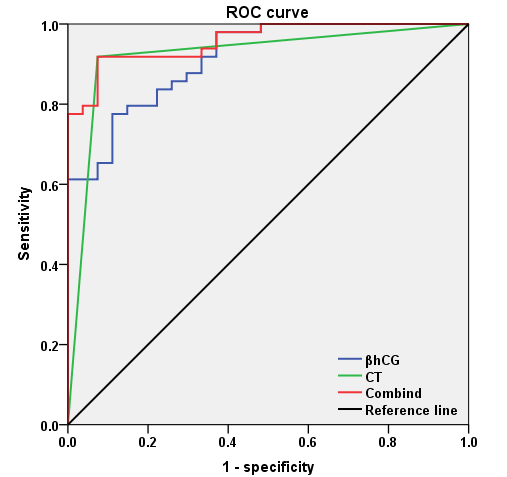
Figure 1 ROC curves of three-dimensional esophageal CT, serum β-hCG, and their joint detection.
4 Discussion
Due to the complicated pathogenesis of esophageal cancer and unclear etiology, a single indicator cannot make a clear diagnosis of the disease, so multiple indicators need to be jointly used to improve its diagnostic level [16]. In order to improve the diagnosis of esophageal cancer, and ameliorate the efficacy and prognosis of patients, this paper explored the application value of three-dimensional esophageal CT combined with serum β-hCG in esophageal cancer. Our results suggested that three-dimensional esophageal CT combined with β-hCG may have a better application value in the diagnosis of esophageal cancer.
The accuracy and density resolution of CT imaging diagnosis are high, which can show the size, metastasis path, diffusion range, and surrounding infiltration of the esophageal mass, and have certain advantages in the diagnosis of esophageal cancer [17]. The outcomes of this study revealed that three-dimensional esophageal CT has a high accuracy rate in the diagnosis of esophageal cancer. Compared with two-dimensional imaging, three-dimensional esophageal CT can demonstrate the overall morphological changes of the esophagus by intuitively presenting stereoscopic three-dimensional images, clearly show the lymphatic metastasis of each region and the infiltration of surrounding tissues and provide a more accurate structure of the tumor, which is contributive to performing more accurate preoperative staging, as well as assessing and predicting the total area of the tumor to select optimal surgical treatment plan [18,19]. When performing diagnostic examination of esophageal cancer, the metastasis of esophageal cancer in lungs, chest wall and liver can also be observed due to the wide scanning area [19]. Therefore, three-dimensional esophageal CT has a better application value in the diagnosis of esophageal cancer.
β-hCG is a hormone produced by tumor syncytiotrophoblast cells whose level in serum or urine will be increased in patients with some non-trophoblastic malignant tumor diseases such as testicular cancer, embryoma, and cervical carcinoma [12]. Accordingly, β-hCG is widely used in clinical practice for the diagnosis of cervical carcinoma and ovarian tumor [12,20]. This study found that serum β-hCG level in patients with esophageal cancer was higher than that in benign patients, indicating that β-hCG level is associated with the occurrence and development of the disease and can be used as a diagnostic basis for esophageal cancer. The possible reason for this is that serum β-hCG is a glycoprotein synthesized and secreted by tumor syncytiotrophoblast cells, and patients with esophageal cancer have weaker immune function and higher risk of infection as compared with normal people or benign patients due to the influences of tumors, which leads to rapid cell proliferation and production of a large number of β-hCG, resulting in the elevation of β-hCG level in the bodies [21]. Hence, serum β-hCG can be used as an auxiliary diagnostic indicator for esophageal cancer.
Moreover, gastroscopic biopsy, three-dimensional esophageal CT, and serum β-hCG examination were performed in all patients with suspected esophageal cancer, and the detection rates of different diagnostic methods were compared. The results uncovered that the diagnostic accuracy of three-dimensional esophageal CT combined with β-hCG examination was high. Three-dimensional esophageal CT could clearly show the morphology of the tumor in the esophagus and the involvement of tissues and organs, and lymphatic metastasis through the secondary analysis of the data, which has a certain value in the diagnosis of esophageal cancer [21]. When combined with β-hCG examination, it has high accuracy, sensitivity, and specificity, with an application value higher than a single diagnostic index, which can reduce the under-diagnosis rate of esophageal cancer [21]. Therefore, three-dimensional esophageal CT combined with β-hCG examination has high diagnostic value for patients with esophageal cancer. However, in this study, due to the limited number of sample cases and review time, the results were not sufficient to represent all patients' conditions, and the diagnosis of three-dimensional esophageal CT combined with β-hCG examination needs further investigation to improve the relevant theoretical research.
In conclusion, three-dimensional esophageal CT combined with serum β-hCG may have a better diagnostic value in esophageal cancer.
Back Matter
Acknowledgments
Not applicable.
Conflicts of Interest
The authors declare no conflicts of interest.
Author Contributions
Data curation, Formal analysis and Methodology, W.S.; Conceptualization, Writing - Original draft, Writing - review and editing, Y.Z. All authors have read and agreed to the published version of the manuscript.
Ethics Approval and Consent to Participate
The study was approved by Medical Ethics Committee, and the patients were informed and consented.
Funding
This research received no external funding.
Availability of Data and Materials
The data presented in this study are available on request from the corresponding author.
Supplementary Materials
Not applicable.
References
- Watanabe M, Otake R, Kozuki R, et al. Recent progress in multidisciplinary treatment for patients with esophageal cancer. Surgery Today 2020; 50(1): 12-20.
- Takeuchi H, Miyata H, Gotoh M, et al. A risk model for esophagectomy using data of 5354 patients included in a Japanese nationwide web-based database. Annals of Surgery 2014; 260(2): 259-266.
- Huang FL, Yu SJ. Esophageal cancer: Risk factors, genetic association, and treatment. Asian Journal of Surgery 2018; 41(3): 210-215.
- Elliott JA, Docherty NG, Eckhardt HG, et al. Weight Loss, Satiety, and the Postprandial Gut Hormone Response After Esophagectomy: A Prospective Study. Annals of Surgery 2017; 266(1): 82-90.
- Visaggi P, Barberio B, Ghisa M, et al. Modern diagnosis of early esophageal cancer: from blood biomarkers to advanced endoscopy and artificial intelligence. Cancers 2021; 13(13): 3162.
- Mönig S, Chevallay M, Niclauss N, et al. Early esophageal cancer: the significance of surgery, endoscopy, and chemoradiation. Annals of the New York Academy of Sciences 2018; 1434(1): 115-123.
- Ishihara R, Goda K, Oyama T. Endoscopic diagnosis and treatment of esophageal adenocarcinoma: introduction of Japan Esophageal Society classification of Barrett's esophagus. Journal of Gastroenterology 2019; 54(1): 1-9.
- Li D, Zhang L, Liu Y, et al. Specific DNA methylation markers in the diagnosis and prognosis of esophageal cancer. Aging 2019; 11(23): 11640-11658.
- Li L, Wang S, Zheng J, et al. Clinical value of upper gastrointestinal radiography and three-dimensional esophageal CT in the diagnosis of esophageal cancer. Journal of Medical Imaging 2023; 33(4): 693-696.
- Liu K, Zhao T, Wang J, et al. Etiology, cancer stem cells and potential diagnostic biomarkers for esophageal cancer. Cancer Letters 2019; 458: 21-28.
- Brancato V, Garbino N, Mannelli L, et al. Impact of radiogenomics in esophageal cancer on clinical outcomes: A pilot study. World Journal of Gastroenterology 2021; 27(36): 6110.
- Li H, Li C, Gong J. Diagnostic significance of serum alpha-fetoprotein combined with β-hCG level in nonseminomatous germ cell tumor. Health Vocational Education 2018; 36(17): 155-157.
- Singh J, Swaminathan U, Sharada P, et al. Estimation of expression of beta-human chorionic gonadotropin levels through progression of disease from normal to epithelial dysplasia to malignancy. Journal of Oral and Maxillofacial Pathology: JOMFP 2019; 23(1): 108.
- Kang S, Zaidi A J, Shokouh-Amiri M, et al. A case report of paraneoplastic syndrome in β-hCG-secreting duodenal adenocarcinoma. Journal of Gastrointestinal Oncology 2019; 10(6): 1151.
- Bagaria B, Bagaria A, Singh M, et al. Diagnostic sensitivity of serum carcinoembryonic antigen, carbohydrate antigen 19–9, alpha-fetoprotein, and beta-human chorionic gonadotropin in esophageal carcinoma (receiver operating characteristic curve analysis). Clinical Cancer Investigation Journal 2015; 4(3): 312-317.
- Zheng R, He S, Zhu H, et al. Diagnostic value of CYFRA21-1, CA19-9 and β-hCG in patients with esophageal cancer and its correlation with T lymphocyte subsets. Journal of Modern Medical Laboratory 2021; 36(1): 51-53+91.
- Liang S. Barium contrast and CT diagnostic value of esophageal cancer. Imaging Research and Medical Application 2021; 5(10): 161-162.
- Yue Y, Li N, Shahid H, et al. Gross Tumor Volume Definition and Comparative Assessment for Esophageal Squamous Cell Carcinoma From 3D 18F-FDG PET/CT by Deep Learning-Based Method. Frontiers in Oncology 2022; 12: 799207.
- Geng Y, Ren Y, Li Z, et al. The value of CT iterative reconstruction in preoperative evaluation of hilar cholangiocarcinoma resection. Progress in Modern Chinese General Surgery 2022; 25(6): 467-469.
- Dinis de Sousa M, Barata M, Miranda AR, et al. Beta-HCG secretion by a pulmonary pleomorphic carcinoma: A case report. Respiratory Medicine Case Reports 2021; 34: 101528.
- Gao Y, Gong C, Wang H, et al. Clinical value of combined examination of serum β-HCG PCT and CRP and secretion GBS in predicting chorioamnionitis and neonatal infection in pregnant women with premature rupture of membranes. Hebei Medical Journal 2021; 27(6): 930-934.